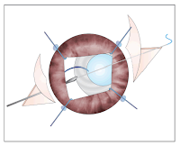
Treatment options for symptomatic
subluxated or dislocated IOLs include observation; and repositioning, removal,
or exchange of the IOL. We describe a surgical technique of trans-scleralsuture fixation for subluxated bag-IOL complex. A 29 year old male; known case
of bilateral recurrent tubercular panuveitis underwent left eye
phacoemulsification with three piece hydrophobic acrylic IOL implantation a
decade ago.
Monday, 24 April 2017
Friday, 21 April 2017
General on Glaucoma and Oxidative Stress. Comments on Study Design: “Biomarkers of Lipid Peroxidation in the Aqueous Humor of Primary Open angle Glaucoma Patients”
Glaucoma is an optic neuropathy
that causes progressive changes in the visual field and whose main known risk
factor is the increased IOP. It is true that the latest acquisitions in image analysis technology (Optical Coherence Tomography -OCT-) have provided objective and quantifiable data of morphological damage, in any way eliminates
the subjectivity and variability of the methods previously employed. If we
speak from the functional point of view, computerized perimetry remains the
method most commonly used scanning glaucomatous damage worldwide. While
exploring the optic disc remains the way easier to assess the damage to the
optic nerve, the great variability in the interpretation and errors derivatives
thereof, the OCT has become critical in monitoring patients with glaucoma.
Wednesday, 19 April 2017
Hypotony as a Hazard of Trabeculectomy with Mitomycin C
An eighty two year old Caucasian
lady with primary open angle glaucoma attended eye clinic. She was using
guttate latanoprost 50 g/ml, Brimonidine 2 mg/ml and combined Dorzolamide 20
g/ml and Timolol 5 mg/ml.
This lady was myopic with right eye
manifest refraction spherical equivalent of -2.00 and left eye manifest
refraction spherical equivalent of -8.00 dioptres. The left eye was amblyopic as a result of this anisometropia. This lady had had bilateral uncomplicated
cataract extractions by phacoemulsification with intraocular lens implantation
and subsequently bilateral neodymium yttrium aluminium garnet or Nd: YAG laser
posterior capsulotomies.
This patient had a medical history
of pulmonary tuberculosis and pulmonary fibrosis. She also had aortic valve regurgitation and osteoarthritis. Her regular medications were Digoxin 125 μg,
Bumetanide 1mg and Lisinopril 1mg daily. She utilized supplementary oxygen for
16 hours daily.
Tuesday, 18 April 2017
Combined Antifungal therapy is effective in handling fungal keratitis
Fungal keratitis is an alarming
disease with a significant impact on the vision quality. Although anti fungal drugs are effective in controlling this disorder, a significant challenge lies
in the management of the disease.
The drug delivery into the corneal tissues
and identification of fungal pathogens play important roles in management of
fungal keratitis. Although 10 different varieties of antibacterial, antifungal
and cycloplegic drugs are available, a combined therapy of antifungal agents
achieved the best treatment modality in cases of fungal keratitis.
Monday, 17 April 2017
Intraocular Pressure Measurement after Photorefractive Keratectomy: Does Contact Area Matter?
Refractive laser surgery induces
substantial changes in corneal structure, causing inaccurate intraocular
pressure (IOP) readings. Pascal dynamic contour tonometry (PDCT) and I care rebound tonometer (RBT) are two novel devices that do not depend on applanationto measure IOP. Purpose of this prospective study was to compare PDCT and
rebound tonometry versus Gold man tonometry (GAT) in a group of patients who
underwent photorefractive keratectomy (PRK).
Central corneal thickness
and IOP were measured in 54 eyes before and after PRK. All IOP measurements
were taken by the same examiner, using PDCT, RBT and GAT in a randomised,
masked fashion. After excimer laser surgery, PDCT measurements were higher than GAT (p<0.0001) and RBT (p=0.0012). Multiple linear regression
analysis indicated that size of contact area was significant (b=-0.504;
p<0.0001) while corneal thickness was not (b=0.003; p=0.169).
Monday, 10 April 2017
Balancing Patient and Practitioner Goals in Contact Lens Fitting
Patients generally are seeking
three things from their contact lens wearing experience, vision, comfort, and
convenience. In addition, some individuals will be looking to seek these goals
with a minimum of “out of pocket” expense. As an eye care provider our goal in contact lens fitting is to provide for optimal ocular health while also establishing a vision correction modality that maximizes clear binocular vision. However,
there may be situations when we need to strike a balance between what patients
can and will do, as contrasted with what might otherwise seem the ideal
situation.
There is a wealth of published data
that clearly indicates that patients do not always replace their lenses with
the frequency we would like. Sometimes this is due to negligence, but often the underlying motivation is simply cost. We need to remind ourselves that the
replacement period for most contact lenses is not dictated by the FDA, but is
based on recommendations made by the manufacturer. The following is taken from
the package insert of a commonly prescribed silicone hydrogel lens.
Tuesday, 4 April 2017
Surgical Management of Glaucoma in Sturge-Weber Syndrome
Glaucoma is a common feature, with
an incidence of 30%-71% in patients with Sturge-Weber syndrome. Many mechanisms
of raised intraocular pressure have been described in the past, the most consistent being congenital trabeculodysgenesis, increased episcleral venous pressure and hypersecretion due to ciliary body angioma.
An increased risk of intra and
post-operative complications has been noted with glaucoma filtering procedures
in these patients, predominantly due to rupture of the fragile vasculature in
the choroidal hemangiomas, leading to expulsive choroidal haemorrhage or
exudative choroidal detachment (CD) caused by sudden decompression during or
after filtering procedures. Prohylactic sclerotomies have been advocated, to be
performed prior to ocular decompression, during filtering procedures in order
to avoid these complications. The necessity of prophylactic procedures has been questioned. Eibschitz-Tsimhoni et al., in a retrospective study, have reported
that none of their 17 patients with SWS who underwent glaucoma filtering
surgery without prophylactic posterior sclerotomy developed intraoperative
suprachoroidal haemorrhage or choroidal effusion requiring therapeutic
intervention.
Monday, 3 April 2017
Saints and Ophthalmology: A Pattern for Emulation, a Model of Healing, and Physicians in Action
From the earliest times, medical
practitioners have sought divine help and support to aid them as they go about
their busy rounds. In Christian Europe of the High Middle Ages, saints played a central role in the everyday life of the ailing. Alongside healing attempts
which involved magic, folk or scientifically-based medicine, the invocation of
specific patron saints for the curing of ailments was a widespread practice.
The miracles of vision performed by saints over the past two millennia are
listed and interpreted by various reviewers along faith healing, spontaneous
recovery, or physicians acting as saints. The individuals honored as patron saints of medicine were practicing physicians who served their patients and their communities well. It is their role as saints to represent the spiritual
element in the healing process as well as personifying the charitable idealism
of the good physician, and therefore are models for humanitarian physicians,
with the ultimate model of healing being Jesus Christ.
Subscribe to:
Comments (Atom)


