Wednesday, 31 August 2016

Ergonomic Challenges of Employees Using Computers at Work in a Tertiary Institution in Ghana

Ergonomically designed workstationshave direct bearing on the comfort and safety of office computer users. Tremendous usage of computers in most offices of emerging economies have however, not seen accompanying applications of ergonomics in the design of computer workstations despite the numerous benefits. 

Ergonomic Challenges of Employees
Injuries and discomforts therefore have higher propensity to occur since most offices formally designed for paperbased work now accommodate computer workstations, without corresponding redesigning. The study therefore sought to assess computer workstation designs in administrative offices at Kwame Nkrumah University ofScience and Technology, with the aim of creating awareness of ergonomics and its application among administrative office computer users.

A total of 150 office employees purposively sampled participated in this study.Respondents selected included secretaries, research assistants and data and account processors. 

Tuesday, 30 August 2016

A Novel Agent in Retinal Ischemia-Reperfusion Injury

Objective: To investigate the protective effect of recombinant human IL-11 (rhIL-11) administered in a systemic manner on retina tissue during ischemia/reperfusion (I/R) injury in a guinea pig model.

Retinal Ischemia
Study design/materials and methods: An experimental study in retinal I/R. Placebo, ischemia/sham, ischemia/ rhIL-11 groups including five animals in each were formed from male albino guinea pigs. Retinal ischemia was induced by cannulating the anterior chambers and lifting the bottle to a height of 205 cm for 90 Min in the sham and ischemia/rhIL-11 groups. The ischemia/sham and ischemia/rhIL-11 groups received 0.1 cc of asaline solution and 5 μg/kg/day rhIL-11 intraperitoneally one hour before the ischemic insult and during two days of reperfusion, respectively. The guinea pigs were sacrificed for biochemical analysis and the levels of tumor necrosis factor alpha (TNF-α), transforming growth factor beta (TGF-β) and interleukin-6 (IL-6) in retina were analyzed with ELISA. Mann–Whitney-U and Kruskal–Wallis tests were used for statistical analysis.

Monday, 29 August 2016

Amantadine Corneal Toxicity: Case Report and Review of the Literature

To report a case of bilateral corneal edema in a patient with a history of cocaine abuse and bipolar-depression treated with amantadine for 6 months, and to review the literature on amantadine corneal toxicity.

The clinical record of a patient with amantadine corneal toxicity and the existing literature on this subject were extensively reviewed for prevalence, clinical presentation, pathogenesis and therapy.

Amantadine Corneal Toxicity
A 27 year-old male who suffered from depression and cocaine abuse presented with sudden visual loss (20/150 OD and 20/200 OS) caused by bilateral corneal stromal edema without ocular inflammation. Corneal edema was related to amantadine therapy (200 mg per day) for 6 months. Visual acuity improved to 20/20 OU and corneal edema resolved after one month of cessation of therapy. Corneal endothelial cell density after3 months of discontinuation of amantadine therapy was 636 ± 359 cells/mm² in the right eye and 1,176 ± 238 cells/mm² in the left eye. There was also increased pleomorphism (45.3% 6A cells OD, 56.4% 6A cells OS) and polymegatism (coefficient of variation, 39.04 ± 6.38 OD and 34.76 ± 2.68 OS).

Thursday, 25 August 2016

Correlation of Retinal Vessel Analysis and Nerve Fiber Layer Thickness in Normal Tension Glaucoma

Background: To investigate the retinal vessel diameter and its possible relationship with the retinal nerve fiber layer (RNFL) thickness in normal tension glaucoma (NTG).

Methods: 86 NTG patients with detailed eye examination including retinal vessel analysis (DVA) and measurement of the RNFL thickness with scanning laser polarimetry (SLP; GDxVCC) were included in this retrospective study. The temporal retinal arteriolar diameters, temporal retinal venular diameters, vascular flicker response were compared with RNFL thickness and NFI (nerve fiber index).

Dynamic vessel analysis
Results:The flicker response of DVA was significantly reduced as compared to normal eyes. The diameters of the temporal retinal arteriolar vessels were significantly correlated with RNFL thickness (P=0.0204) and NFI of GDxVCC (P=0.0021). The diameters of the temporal retinal venular were significantly correlated withthe NFI (P=0.0298).
Conclusion: Our results show that narrower arteriolar vessels are found in advanced NTG patients. These findings may be due to the decreased demand for retinal blood flow in damaged retina, but also may be one reason for the damage. Vessel analysis can contribute to the available tools predicting glaucomatous damage before visual field loss becomes manifest.

Wednesday, 24 August 2016

Evaluation for Dry Eye and Intra Ocular Pressure in Patients on Antihypertensive Medications

The study was conducted to evaluate and analyze the ocular side effects of antihypertensive drugs in the patients and to determine the effects of antihypertensive drugs in causing dry eye disease and intra ocular pressure changes. This was a case-control, non-interventional study of 140 patients (70 cases and 70 controls) conducted from June 2014 to May 2015 in Dr. Rajendra Prasad Government Medical College, Kangra at Tanda, Himachal Pradesh (India). 

Dry Eye
Patients were subjected to detailed history and examination using standardized protocol and Performa. The prevalence of dry eye in this study was 44.3% in cases and 7.1% in controls, with p value of 0.0001 (<0.05), which is significant. Dry eye was present in allthe patients on beta blockers and thiazide diuretics (alone or withcombination). Dry eye disease was significantly associated with increasing age and longer duration of antihypertensive treatment. It was observed that average IOP was lower in patients on antihypertensive treatment as compared to normal subjects.

Tuesday, 23 August 2016

Vision-related Quality of Life in Children with Amblyopia

Vision plays an important role in most everyday activities. Consistent with this, people with visual impairment are usually faced with significant challenges in their daily activities. In children, such activitiesinclude playing, reading, socialisation and taking care of their daily needs. 

Vision-related QualityIn the paediatric ophthalmological field, visual problems include high refractive errors, binocular disorders, depth perception deficiency, amblyopia and ocular pathology. These visual impairments in children potentially causepsychological and functional changes and could affect educational and social prospects and may thus impact on vision-related quality of life (VRQoL).
Amblyopia is usually defined as a unilateral or bilateral reduction in visual function caused by abnormal visual input resulting from degradation of the retinal image during a sensitive period of visual development, which historically has been thought to be the first seven years oflife.

Monday, 22 August 2016

B-Scan Dynamic Sonographic Studies in Orbital Pathologies

orbital Pathologies impact factor
For pretty long time, the ophthalmologist used to establish the diagnosis of eye diseases on clinical ground, slit lamp examination and fundus examination before medical or surgical treatment. Ultrasonography study using 10 - 12 MHz small probe did wonder in the field of eye and orbital disease. A stands for amplitude or time scan whereas B stands for brightness scan. Both are used for visualization and measurement of ocular pathology and anatomy.Read More...

Friday, 19 August 2016

Use of the American Academy of Ophthalmology Surgical Checklist and Implant Timeouts

Surgery has become an important component of healthcare, but along with recognition of its ability to enhance public health is growing attention to its potential for substantial harm if practiced unsafely. In light of this, the World Health Organization (WHO) established the Safe Surgery Saves Lives initiative in 2007 as the second of their Global Patient Safety Challenge topics. 


This initiative promulgated routine use of a surgical checklist to ensure systematic adherence to steps designed to promote safe surgical practice. The Safe Surgery Saves Lives initiative led to an oft-cited 8-city prospective trial of a 19-item checklist, which showed reductions in complications and deaths after intervention – a result that has since been reproduced by studies in other countries and practice settings.

Surgical checklist


The idea that surgical error rates can be improved by actionable changes to a system that facilitates errors is of particular relevance to the field of ophthalmology. First, ophthalmology accounts for a significant proportion of surgical volume. 

Cataract surgery alone is the most commonly performed surgical procedure in the United States Medicare population, and is projected to increase in the future. Second, there is ample evidence of surgical errors in ophthalmology, referred to by the literature variously as “surgical confusions”, “never events”, and “sentinel events”.Read More...

Thursday, 18 August 2016

Balancing Patient and Practitioner Goals in Contact Lens Fitting

Patients generally are seeking three things from their contact lens wearing experience, vision, comfort, and convenience. In addition, some individuals will be looking to seek these goals with a minimum of “out of pocket” expense. As an eye care provider our goal in contact lens fitting is to provide for optimal ocular health while also establishing a vision correction modality that maximizes clear binocular vision. 


There is a wealth of published data that clearly indicates that patients do not always replace their lenses with the frequency we would like. Sometimes this is due to negligence, but often the underlying motivation is simply cost. We need to remind ourselves that the replacement period for most contact lenses is not dictated by the FDA, but is based on recommendations made by the manufacturer. The following is taken from the package insert of a commonly prescribed silicone hydrogel lens.

Balancing Patient

“When prescribed for daily wear (frequent replacement), it is recommended that the lenses be discarded and replaced with a new lens every 2 weeks. However, the Eye Care Professional is encouraged to determine an appropriate replacement schedule based upon the response of the patient.”Read More....

In-toto Removal of Sub- Retinal Cysticercosis and its Histopathology

To study the epidemiology and clinical aspects of the hypertensive retinopathy among pregnant women with hypertension in the CHUD-B. Material and methods: The study was multicenter and analytical. It concerned all pregnant women admitted to the CHDU-B from April 1st to July 31, 2012, four months. 

Results: Out of 1327 pregnant women, 158 (11.90%) have presented an arterial hypertension associated with pregnancy. Among these 158 pregnant women with hypertension, 112 have benefited from the ophthalmological examination, for a participation rate of 70.88%. 48 had retinopathy hypertensive either a prevalence of 42.86%. The average age of pregnant women was 28 ± 7.76 years. 

Sub retinal cysticercus

These women were in the majority illiterate (38%) with a predominance of craftswomen and housewives (66.97% ). The HTA was severe in 54.17% of cases and the hypertensive retinopathy was statistically linked to the severity and chronicity of the HTA. Conclusion: The hypertensive retinopathy during pregnancy is common and affects women around the thirties. It is often related to the severity and chronicity of the HTA.Read More....

Wednesday, 17 August 2016

A Decade of Anti-VEGF Drugs in Ophthalmology- Successes and Challenges

The discovery of anti-VEGF drugs has revolutionized the management of age-related macular degeneration (AMD), diabetic macular edema (DME) and retinal vein occlusion (RVO). We look back to almost ten years of anti-VEGF experience in ophthalmology and remain very optimistic about the therapeutic yield in medical conditions that, before the anti-VEGF era, had a poor long-term prognosis. 
Anti-VEGF treatment

At those times, where photodynamic therapy in combination with verteporfin was the only principled approach in AMD management, ophthalmologists had usually bad news for their patients and actually accompanied them to the state of complete loss of central vision.
Today patients are promised a clear treatment response if anti- VEGF medications are provided timely. A network meta-analysis, comparing the two currently licensed drugs Ranibizumab (Lucentis®) and Aflibercept (Eylea®) against placebo, showed that the benefits for all relevant efficacy outcomes were consistently positive. Read More.....

Tuesday, 16 August 2016

Paediatric Aphakic Glaucoma: A Diagnostic and Management Challenge

Pediatric aphakic glaucoma is a potentially blinding condition, which poses significant management challenges - the patient is young and treatment must aim to preserve vision for perhaps 80 years or more of life expectancy. The purpose of this study is to highlight that early detection is important to improve the prognosis for these patients and their families. Misdiagnosis of this condition may adversely affect vision preservation. 


However, diagnosis can be difficult, especially when children may demonstrate vague symptoms such as irritability, photophobia and epiphora which themselves may further impair clinical co-operation. Despite considerable advances in techniques employed in paediatric cataract surgery, aphakic glaucoma continues to occur and remains notoriously challenging to manage. 

Paediatric Aphakic Glaucoma

We present a case of glaucoma after cataract surgery that masqueraded as corneal infection which was challenging to diagnose and treat. Prompt recognition of pediatric aphakic glaucoma and intraocular pressure (IOP) control has resulted in a favorable clinical outcome, thus far.Read More.....

Hypertensive Retinopathy during Pregnancy

To study the epidemiology and clinical aspects of the hypertensive retinopathy among pregnant women with hypertension in the CHUD-B. Material and methods: The study was multicenter and analytical. It concerned all pregnant women admitted to the CHDU-B from April 1st to July 31, 2012, four months.

Results: Out of 1327 pregnant women, 158 (11.90%) have presented an arterial hypertension associated with pregnancy. Among these 158 pregnant women with hypertension, 112 have benefited from the ophthalmological examination, for a participation rate of 70.88%. 48 had retinopathy hypertensive either a prevalence of 42.86%. 

Hypertensive Retinopathy during Pregnancy

The average age of pregnant women was 28 ± 7.76 years. These women were in the majority illiterate (38%) with a predominance of craftswomen and housewives (66.97% ). The HTA was severe in 54.17% of cases and the hypertensive retinopathy was statistically linked to the severity and chronicity of the HTA. Conclusion: The hypertensive retinopathy during pregnancy is common and affects women around the thirties. It is often related to the severity and chronicity of the HTA. Read More...

Thursday, 11 August 2016

The Surgical Outcomes, Complications and Predictive Surgical Factors of Diabetic Retinopathy Vitrectomy

Purpose: To evaluate the visual, anatomical outcomes, complications and the predictive preoperative and intraoperative factors of pars plana vitrectomy (PPV) for proliferative diabetic retinopathy (PDR) in a large Asian tertiary eye center.

Methods: This is a consecutive retrospective review of 106 eyes that underwent PPV for PDR complications in 2013. The functional success was defined as visual acuity of 20/200 or better while the anatomical success was defined as 360° flat retina without endo-tamponade at one year.


Results: The overall functional and anatomical success was 77.4% and 94.3%, respectively. The common complications were iatrogenic break (14.2%), raised intraocular pressure (IOP) (13.2%), cataract (13.2%) and recurrent vitreous hemorrhage (12.3%). The use of intraoperative triamcinolone (OR: 0.36, p=0.04) and silicone oil (OR: 0.08, p<0.001) were associated with poorer visual success. The iatrogenic breaks occurred more often in 20G PPV than 23G PPV (OR: 5.89, p=0.02) whereas intraoperative silicone oil were associated with postoperative raised IOP (OR: 3.71, p=0.04). The use of bevacizumab was not found to reduce recurrent vitreous hemorrhage (OR: 0.53, 95%CI: 0.11-2.53, p=0.43).Read More....

Tuesday, 9 August 2016

Impact of Amblyopia Treatment on Vision-Related Quality Of Life

Treatment of amblyopia during childhood can have both positive and negative impacts in later life. The treatment of amblyopia could influence Vision-related Quality of Life in adults and children, particularly social relationships and emotions. For example, children with a history of occlusion were 35% to 37% more likely than children without visual defects to have suffered from verbal or physical bullying at school. Many parents associate occlusion treatment with a decrease in children’s self-confidence because of poor vision during occlusion and report greater distress and more conflict at home.

Koklanis et al. conducted a study in Australia on the psychosocial impact of amblyopia and its treatment from both the children’s (aged 3 to 15 years) and parents’ perspectives. The investigators performed asemi-structured interview with both children with amblyopia and their parents. In addition, parents were asked to complete a psychological inventory, the Behaviour Assessment System for Children. The study showed that dealing with stigma and the perceptions and responses of peers were found to be of central significance in amblyopia therapy and that stigma and the perceptions of peers had adverse consequences for some children’s identity and psychosocial wellbeing.

Amblyopia Treatment


In contrast, some studies have shown that parents of amblyopic children undergoing occlusion therapy do not report more stress or more psychosocial impacts in their children than parents of children who were not occluded. The level of parent’s stress and child's psychosocial wellbeing in the occluded group did not notably change following the onset of occlusion treatment. For example, in the United Kingdom, Choong et al. investigated the psychosocial impact of occlusion therapy on children and their guardians using a questionnaire of perceived stress index (PSI) and the perceived psychosocial questionnaire (PPQ). Findings from this study showed that carers of children undergoing occlusion therapy did not experience statistically significant additional stress or perceive their child as showing poorer psychosocial wellbeing compared to carers in the non-occluded group. 

In the occluded group, the stress level of guardians and the child’s psychosocial wellbeing did not significantly alter the subsequent onset of occlusion treatment. Likewise, this finding is consistent with the previous finding that parents’ perspectives differ from those of children. This study found no evidence to indicate thatocclusion therapy has negative psychosocial impact on carers and childrenalike. Koklanis et al. suggested that this can occur if the “parents, siblings and peers also assisted in maintaining good self-esteem and a good attitude towards their treatment”. In other words, it is the behaviour of people close to the child with amblyopia that can determine whether or not they suffer any psychological impacts due to social relations and that is often a result of society and culture and the quality of interpersonal relationships.

Detecting the Physiological Blind Spot with Reaction Time Perimeter

Objective: To study the ability of a novel reaction time perimeter to detect a physiological blind spot. Methods: The location of the physiological blind spot of 11 healthy volunteers was determined with two independent methods, first by Octopus custom-made blind spot visual field program, and second by Fundus photography with a method previously described by the authors. With reaction time perimeter saccade triggering stimuli (STS) were shown in predetermined locations of the visual field in order to initiate saccades. 

An STS was followed by an FO (fixation object) i.e., an arrow head pointing either to the right or left at the same location as the previously shown STS. The recognition of the FO was reported by pressing a correct button(right or left respectively) thus verifying accurate fixation by the fovea. Time intervals between button presses were recorded. Reaction times for detecting the STS within the blind spot area were compared to those for detecting the STS in other locations of the visual field. Results: Reaction times within the blind spot area were longer compared to those in other locations in 10 of 11 subjects (Analysis of variance), p<0.001 in 9 subjects, p<0.006 in one subject). Conclusion: A physiological blind spot can be detected with reaction time perimeter.
Physiological Blind Spot

The ability to move the eyes towards an object perceived in the peripheral visual field in order to fixate with the fovea is a fundamental feature of the visual system. Visual processing speed, which is defined as the time needed to make a correct judgment about a visual stimulus, is commonly studied in behavioral research by measuring reaction times. Visual field defects can be quantified by standard automated perimetry (SAP), but it has limited power to predict the impact of disease on the ability to perform activities of daily living. The testing situation in SAP involves maintaining a steady fixation whilst keeping the head still on a head rest making it very unnatural compared to real-world viewing tasks.

In this pilot study, our purpose was to determine if reaction time perimeter allowing free head and eye movement and thus simulating the normal features of the visual system can be used to detect a physiological blind spot.


Monday, 8 August 2016

Prevention of Infectious Keratitis after Corneal Refractive Surgery

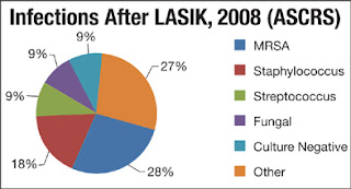
Corneal infections are, although not frequent, a major concern after laser in situ keratomileusis (LASIK) and surface ablation (SA) techniques (PRK, LASEK and epi-LASIK).

The real prevalence of infectious keratitis after refractive surgery is difficult to determine, due to its rarity. In 2010 we reported a prevalence of 72 cases in 204,586 LASIK procedures, a 0.035%. Other series had shown prevalences ranging from 0.034 to 0.180%, but ours was, by far, the largest series published to date. As happens in LASIK, the prevalence of infections after SA is rare and it is difficult to determine if the series does not comprise a large number of operated eyes. In 2011 we published our prevalence in SA. We found 39 cases in 18,651 procedures, a 0.200%. Therefore, in our institution, corneal infections were five times more prevalent after SAthan after LASIK.

Prevention of Infectious Keratitis after Corneal Refractive Surgery

There is much consensus about the standard treatment of corneal infections after corneal refractive surgery. According to the ASCRS White Paper aggressive management with early flap lifting, scraping, culture, and irrigation with antibiotics should be followed. When the infection has an early onset (less than 15 days) moxifloxacin 0.5% (or gatifloxacin 0.3%) is recommended along with cefazolin 50 mg/ml every 30 minutes. If it has a late onset (more than 15 days) moxifloxacin 0.5% (or gatifloxacin 0.3%) must be alternated with amikacin 35 mg/ml. In every case, oral doxicyclin is to be administered (100 mg, twice a day).