Tamoxifen is a drug used to treat
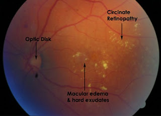
estrogen receptor positive breast cancer that can induce retinopathy. The diagnosis of tamoxifen retinopathy is traditionally established by macularedema seen on fluorescein angiography and retinal crystalline deposits seen on funduscopy. Macular edema associated with tamoxifen retinopathy has been
reported to be reversible after cessation of the drug but the retinal crystalline
opacities usually persist.
Recently, a case of bilateral microcystoid
maculopathy with patches of photoreceptor loss associated with concurrent
tamoxifen use was detected using a research-grade high resolution
Fourier-domain optical coherence tomography (Fd-OCT) in a patient with vision
loss unexplained by funduscopy, fluorescein angiogram (FA), multifocal electro retinography and Stratus OCT. This report describes two new cases of maculopathy associated
with prior tamoxifen use in which similar morphologic changes were seen using
commercially available Fd-OCTs, Cirrus (Carl Zeiss Meditec, Dublin, CA) and
RTVue (Optovue, Fremont, CA), in eyes that appeared unremarkable on funduscopy.

No comments:
Post a Comment