Friday 30 September 2016

Focal Necrosis in Orbital Pleomorphic Adenoma

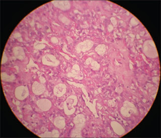
Pleomorphic adenomas (PA) are common benign tumours of the lacrimal gland. Necrosis in PA is unusual and should raise a suspicion and screen for malignancy. We hereby present a casereport of pleomorphic adenoma with necrotic foci in the orbit of a 44-year-oldlady and a review of current literature for focal necrosis in orbitalpleomorphic adenoma.

Focal Necrosis
Lacrimal gland tumours are rare entities that only comprise 9% of orbital lesions. Of those 9% of cases, only 10% are pleomorphic adenomas. Pleomorphic adenomas (PA) are the most common benigntumours of the lacrimal gland, which exhibit pleomorphism of epithelialcomponents. However there are certain histological characteristics in pleomorphic adenomas, which may raise suspicion of atypia and prompt further investigation. Auclair and Ellis ET al. has noted that areas of necrosis, hyper cellularity, hyalinization, cytological atypia, capsule extension or violation may be predictors of malignancy.
Thursday 29 September 2016

Correlation of Tono-Pen and Pneumatonometer Measurements from Four Scleral Quadrants with Intraocular Pressure Measurements from the Cornea

To assess whether Tono-Pen and pneumatonometer measurements obtained from the sclera correlate to intraocular pressure measurements obtained from the cornea. This is a prospective, cross-sectional study,conducted at the New York Eye and Ear Infirmary of Mount Sinai. Patients wererandomized to have their left or right eye included in the study. 

Tono-Pen
Exclusion criteria included prior intraocular surgery (except uncomplicated cataract extraction), uveitis, corneal or scleral thinning, central corneal thickness <500 µm or >575 µm, and hyperopia greater than +2 diopters or myopia greater than -4 diopters. Goldmann applanation tonometry, Tono-Pen and pneumatonometer measurements were obtained from the cornea. Tono-Pen and pneumatonometer measurements were obtained from the sclera in the superonasal, superotemporal, inferonasal, and inferotemporal quadrants. Product moment correlation and linear regression were used to examine correlations between the measurements.

Wednesday 28 September 2016

Mini Monoka Stent Insertion for the Management of Epiphora Secondary to Canalicular Obstruction or Stenosis

Epiphora is a common presenting problem in ophthalmological clinical practice. There are multiple causes for epiphora. Canalicular obstruction/stenosis is common and increases with age with a female preponderance. Treatment for epiphora is often controversial and notalways successful. The gold standard of treatment involves retrograde silicone intubation most commonly with Lester Jones bypass tubes plus or minus dacryocystorhinostomy (DCR) or sometimes more complex procedures such as canaliculodacryocystorhinostomy or conjunctivodacryocystorhinostomy if there is total canalicar obstruction. 

Mini Monoka Stent
DCR with retrograde intubation is reported to alleviate or reduces epiphora in 60-70% of patients with proximal or mid-canalicular disease. There is a poorer prognosis for more distal obstructions, with successin the range of 50%. Other complex procedures such as canalicular trephinationwith silicone stent intubation, balloon canaliculoplasty and endocanalicular laser surgery have also recently been described with varying success rates (43 to 84%). Finally, canaliculodacryocystorhinostomy has a reported success rate of approximately 80% if there is common canalicular obstruction whilst conjunctivodacryocystorhinostomy has success rates of around 90% if performed correctly, with unfortunately high complication rates (as high as 50%).

Tuesday 27 September 2016

Rheumatoid keratolysis a silent complication associated with Rheumatoid arthritis

Rheumatoid keratolysis
A 34 year old women suffering from rheumatoid arthritis since 20 years had a complaint of blurred vision in right as she had a problem of corneal melting.  It has been stabilised bytreatment followed by regular monitoring that arthritis was in-active on etanercept.But right cornea revealed 3mm descemetocle, later tectonic corneal transplantation was done and was prescribed tacrolimus. The histological examinations done after three years was characterised by complete loss of corneal stroma due to rheumatoid keratolysis. It has been indicated that metalloproteinase are active in RA are involved in Keratolysis.

Monday 26 September 2016

Neuronavigational Approach for Orbital Neurofibroma Excision: A Case Report

Orbital neurofibromas are uncommon in adults, accounting for approximately 1%-3% of all space occupying lesions of the orbit. The complex anatomy of the orbital region, with the pronounced vulnerability of its neurovascular structures, requires particular surgical precautions. Neuronavigation, as a high-tech device for intraoperative safety,represents a valuable option for the confined orbital space. However, the application of neuronavigation in orbital surgery has been rarely reported. The authors present a case report of a 32-year-old female with an isolated localized neurofibroma surgically approached by intraoperative navigation and a review of the literature.

Orbital Neurofibroma Excision
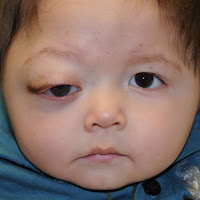
A 32-year-old female presented with a 2-year history of left upper lid swelling and progressive onset of proptosis and double vision (Figure 1). She had no family history of neurofibromatosis.


Ophthalmologist’s examinationrevealed a decreased visual acuity 7/10 associated with ptosis, unilateral exophthalmos with restricted eyeball movement on upward and lateral gaze in the left eye. The visual field examination was normal.

Friday 23 September 2016

A V235L Mutation of the Human BEST1 Gene Associated with Best Macular Dystrophy with Intra-familial Clinical Variations

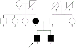
Best vitelliform macular dystrophy (BVMD) accounts for 1% of all cases of macular degeneration resulting in progressive loss of central vision. In this work wesought to evaluate the clinical and genetic background in a two generationpedigree of autosomal dominant BVMD for clinical management and follow up. To our knowledge this is the first report on association of Bestrophin 1 (BEST1) mutation with BVMD in an Indian family.

Human BEST1 Gene
Complete ophthalmic examination done in a family with a complaint of impaired vision revealed the presence of yellow-orange yolk like lesions in the macula upon fundus examination. Further investigations through EOG revealed decreased Arden ratio. Besides the proband,two other members namely, mother and siblings were also affected in the family.Follow up clinical examination was done after three years to clinically document the progression of the disease, OCT examination was done in addition during follow up studies. Genomic DNA samples of the affected family members showed a sequence variation, c.703 G>T transversion in exon # 6 which results in substitution of valine by polar leucine as V235L.

Prevention of Infectious Keratitis after Corneal Refractive Surgery

The real prevalence of infectious keratitis after refractive surgery is difficult to determine, due to its rarity. In 2010 we reported a prevalence of 72 cases in 204,586 LASIKprocedures, a 0.035%. Other series had shown prevalence’s ranging from 0.034 to 0.180%, but ours was, by far, the largest series published to date. 

Infectious Keratitis
As happens in LASIK, the prevalence of infections after SA is rare and it is difficult to determine if the series does not comprise a large number of operated eyes. In 2011 we published our prevalence in SA. We found 39 cases in 18,651 procedures, a 0.200%. Therefore, in our institution, corneal infections were five times more prevalent after SA than after LASIK.

There is much consensus about the standard treatment of corneal infections after corneal refractive surgery. According to the ASCRS White Paper aggressive management with early flap lifting, scraping, culture, and irrigation with antibiotics should be followed. 

Thursday 22 September 2016

Is it safe to use BAK containing eyewashes?

Eyewash Solution on Corneal Epithelium

Usage of eyewashes has increased drastically for healthy eyes due pollinosis and increase usage of contact lenses. Recently they summarised that usage of eye washes containingpreservative BAK showed some disorders such as, collapse of the mucin layer in cornea and epithelium disorders. They also stated that BAK free ophthalmic solutions are used for the treatment of glaucoma, dry eyes and eye allergies. Eye washes lacking BAK showed reduction in corneal disorder.

Tuesday 20 September 2016

Challenges in the Management of Glaucoma in a Patient with Severe Ocular Surface Disease: A Case Report

Ocular surface disease (OSD) is a significant problem for glaucoma patients worldwide, who will require long-term topical treatment. The risk of OSD increases with age, with a reportedprevalence of up to 60%. 

Glaucoma in a Patient
The severity and burden of OSD also increase with the number of topical anti-glaucoma medications used in treatment due to multiple, daily exposures of the ocular surface (OS) to toxic active compounds in the drug itself, other components of the formulation or preservatives. Thepresentation of OSD varies depending on its severity, but usually includessymptoms of dry eyes, allergy and distorted vision, which can be debilitatingand affect quality of life. The clinical signs of the disease include tear-film instability, toxic kerato conjunctivitis, eyelid abnormalities and allergic manifestations. However, the lack of concordance between symptoms and signs often makes the diagnosis of OSD and assessment of its severity challenging.

Monday 19 September 2016

The Review of Behcet Diseases: A Case of Sub-acute Bilateral Hypopyonic Uveitis after 20 Years of Behcet Diseases

Behcet Diseases
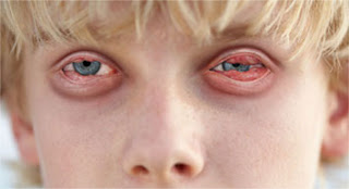
The current study was an attempt to report a case of subacute bilateral hypopionic uveitis and pan uveitis in a referral case of Behcet patient. Careful questioning of the patient anddetailed examination of the eyes for other signs are necessary to guide thedifferential diagnosis and relevant investigations. In this study, a case of a patient with a 20 year history of Behcet disease and occular manifestation of bilateral hypopionic uveitis was presented in ophthalmic consultation. Hypopion resolved shortly after instating therapy but old scarified retinal lesion did not respond as well as long lasting posterior synchia.


Friday 16 September 2016

Focal Necrosis in Orbital Pleomorphic Adenoma

Lacrimal gland tumours are rare entities that only comprise 9% of orbital lesions. Of those 9% of cases, only 10% are pleomorphic adenomas. Pleomorphic adenomas (PA) are the most common benign tumours of the lacrimal gland, which exhibit pleomorphism of epithelialcomponents. However there are certain histological characteristics in pleomorphic adenomas, which may raise suspicion of atypia and prompt further investigation. Auclair and Ellis et al has noted that areas of necrosis, hypercellularity, hyalinization, cytological atypia, capsule extension or violation may be predictors of malignancy.

Automobile Industry


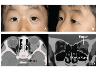
44-year-old Caucasian lady presented with residual painless left upper lid swelling 6 months after being poked in the left eye by her infant son. A firm cystic lesion was palpated in the superior lateral orbital rim. The rest of the ocular examination was normal. Computer Tomographic scan of the orbits revealed a large encapsulatedcystic mass extending from the inferior portion of the left lacrimal gland. A posterior orbitotomy with excisional biopsy of the lesion was performed. This yielded histology of pleomorphic adenoma with cellular myoepithelial areas containing focal necrosis. Cytological atypia and mitotic activity were absent. The patient recovered well post operatively with no complications.

Thursday 15 September 2016

Surgical Management of Macular Hole at 2 Years of Follow-Up

Idiopathic macular holes (MHs) are anatomic defects at the neurosensory central retina extending from the internal limiting layer (ILM) to the retinal pigment epithelium. The physiopathology ofMHs is related to anomalous vitreomacular adhesion in primary MHs. Secondary MHs are rarer and usually related to trauma, laser, or intraocular surgeries. MHs are a significant cause of visual impairment especially in older patients, in whom they develop more frequently. MHs are suspected when patients complain of visual impairment, central scotoma or metamorphopsia and confirmed by fundus examination and optical coherence tomography (OCT).

Macular Hole
Although the original Gass classification is still used widely in clinical practice, a new classification based on OCT findings was reported recently. This classification considers thepresence or absence of vitreomacular traction (VMT), size, and MH etiology. In this OCT-based anatomic classification system, MHs are defined as small when the minimal width is below 250 μm, medium from 250-400 μm, and large over 400 μm. MHs also are classified as having or not having VMT and as primary or secondary forms.

Wednesday 14 September 2016

Contact Lenses: Out with the Old/In with the New?

To answer this question: I certainly hope not, at least not always. Without a doubt over the past several years the contact lens industry has brought forth numerous new materials, designs, and modes of contact lens manufacturing. But that should not translateinto a universal switch from the “old” to the “new”. I would like to share some specific instances where older lens materials and designs should still have a place in your practice.

Contact Lenses
Although without a question newer more oxygen permeable rigid gas permeable (RGP) materials have been key to the introduction of such modalities as overnight wear of Orthokeratology lenses, these materials often have some less advantageous attributes. Although we oftenonly consider oxygen permeability (Dk) and lens wettability when selecting aRGP material, one should also consider that such materials often have lower material hardness and greater modulus. 

Tuesday 13 September 2016

A Clinical Analysis of Age-Related Macular Degeneration in Qinghai Plateau

Age-related macular degeneration (AMD) is a major cause of irreversible vision loss in the elder population of European and American countries. Clinically, AMD has two types: dry AMD (atrophic)and wet AMD (Neovascular or exudative). 80-90% of individuals with AMD are dryAMD. It is characterized by drusen formation on the retina and geographic atrophy in local retinal pigment epithelium (RPE). 
age Related Macular Degeneration

Although wet AMD affects approximately 10-15% of individuals with AMD, it is the direct and major cause of severe vision loss from the disease due to retinal choroid neovascularization (CNV), macular hemorrhage or leakage of fluid into the RPE and subretinal spaces. AMD is a highly complex and multifactorial disease withdemographic, environmental, and genetic risk factors including aging, smoking,diet and race. Thus, the pathogenesis for AMD has yet to be completely understood. In China, AMD has become the third major cause of blindness. A few studies showed a high rate of wet AMD for peoples who lived in Qinghai-Tibet Plateau. However, whether high plateau is a risk factor remains undetermined.

Monday 12 September 2016

Prevention of Recurrence of Ligneous Conjunctivitis with Subconjunctival

Ligneous ConjunctivitisLigneous conjunctivitis (LC) is a rare and chronic disorder which is characterized by hard wood-like membranes on palpebral and bulbar conjunctiva. It usually presents when plasminogenfunctional activity is under normal range (80-120%), usually below 52%, andplasminogen level in blood generally <1.2 mg/dl (normal range 6-25 mg/dl). Severity is associated with a low plasminogen functional activity, values below 5% can affect all mucous surfaces, and the disease could present as a systemic disorder. 

LC may occur after infectious conjunctivitis or ophthalmic surgical treatment, associated with recurrence of membranes after removal. Plasminogen topical administration has demonstrated a plausible efficient treatment for preventing the recurrence of LC. The goal of our study is to report the case ofa 6 year-old male child with post infectious LC, that when he was 2 years old presented recurrence of the disease after surgical resection and combined treatment with topical tobramycin, dexamethasone and cyclosporine A. Treatment with autologous fresh frozen plasma (AFFP) resulted in remission of the membranes.

Friday 9 September 2016

Ischaemic Optic Neuropathy - Arteritic

Ischaemic optic neuropathy (ION) is the commonest adult optic nerve disorder encountered worldwide and can be expected to increase in incidence in our ageing population. The condition hasbeen classified as a) anterior (AION) affecting the optic nerve head and b) posterior (PION) involving that portion of the optic nerve behind its immediate retrolaminar portion. 

Ischaemic Optic Neuropathy
Further more there are two pathological varieties of the disease c) Arteritic (AAION) almost exclusively associated with Giant CellArteritis (GCA) and d) Nonarteritic (NA-AION or less correctly NAION) usually associated with diabetes, hypertension and hypercholesterolaemia. A recent treatise on the subject runs to more than 600 pages.


Thursday 8 September 2016

Unusual Case of an Orbital Lymphangioma Presenting as Chorioretinal Folds

Orbital Lymphangioma
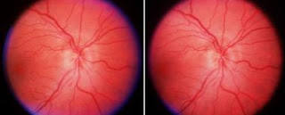
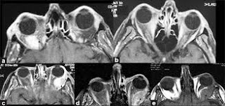
We report a case of 75-year-old elderly male who presented with diminution and distortion of vision, swelling, pain and upward and nasal deviation of eyes. Computed tomography (CT) scan and magnetic resonance imaging (MRI), fundus photography and optical coherence tomography (OCT) were performed. Amultilocular intraconal cystic mass lesion causing chorioretinal folds wasseen. A presumptive diagnosis of orbital lymphangioma with chorioretinal folds was made and was confirmed by histopathology. This case is being reported because of its unusual presentation in an elderly patient (chorioretinal folds with macular involvement).

Wednesday 7 September 2016

Clinical, Functional and Structural Studies of Humans

CDK is a degenerative and potentially handicapping human corneal disease suffered by people living in particular places around the world. This disease ischaracterized by slow progression of corneal opacity. The first description of a corneal disease that may have been CDK was made in 1898 by Baquis  and three decades later by Lugli. 

Structural Studies of Humans
Since then, different descriptions of this disease have been made in different parts of the world characterized by constant winds, low humidity, and chronic exposure to ultraviolet radiation (UVR). Although we have made some significant advancestowards the understanding of CDK pathogenesis over the past few years, we still lack detail of molecular events taking place during CDK onset and progression. The decrease of corneal antioxidant protective mechanisms results in oxidative injury of the cornea and causes damage by UVB rays and by reactive oxygen species generated by them. 

Tuesday 6 September 2016

Anterior Vitreous Incarceration after Phacoemulsification Cataract Extraction

Vitreous incarceration is a condition where vitreous is trapped within a wound or incision site. When involving the cornea, vitreous can prolapse into the anterior chamber and pass through a microscopic wound at the location of an incision. A vitreous strandis visible on slit lamp examination and the condition is often associated witha peaked pupil where the vitreous strand contacts the iris. If the vitreous penetrates through all the corneal layers and onto the extra ocular surface, a vitreous wick syndrome develops significantly increasing the risk for endophthalmitis. Vitreous incarceration can also cause pupillary block glaucoma, cystoid macular edema, vitreoretinal traction, and corneal decompensation.

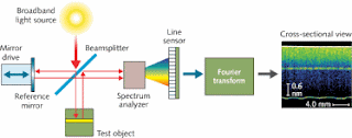
Spectral-Domain Optical Coherence Tomography
Vitreous wick syndrome has been documented as a cause of delayed onset endophthalmitis. In 1970, Ruiz and Teeters described the condition in eleven patients who presented with delayed-onset endophthalmitis after intracapsular cataract extraction surgery (ICCE) with vitreous prolapse. The protruding vitreous strand prevents the wound from closing and can allow micro-organisms to enter. Anterior vitreous wick syndrome is commonly associated with ICCE but has alsobeen reported in cases of extracapsular cataract extraction (ECCE), after posterior capsulotomies, as well as corneal relaxation incisions.

Saturday 3 September 2016

Lacrimal Gland Metastasis and Paraneoplastic Orbital Myositis due to Breast Carcinoma

We present a case with lacrimal gland metastasis and paraneoplasticorbital myositis due to breast carcinoma. A 66 year-old woman with a history of breast carcinoma was seen with the complaints of headache, blurred vision, and diplopia. 

Lacrimal Gland Metastasis
On neuro-ophthalmologic examination restriction of left, right and up gaze in her left eye was found. Magnetic resonance imaging of orbit revealed enlargement of all extraocular muscles, heterogeneous enhancement of lacrimal gland and retroorbital fat in her left eye. The patient underwent biopsy oflacrimal gland and extraocular muscles. Lacrimal gland metastasis was shown by histopathologic examination, but there was no tumor cells in the extraocular muscles. After radiotherapy for lacrimal gland metastasis, her symptoms relieved remarkably. In patients with a history of malignancy, orbital symptoms and findings should be thought as a cause of malignancy primarily.

Thursday 1 September 2016

Microbial Keratitis at an Urban Public Hospital: A 10-Year Update

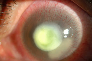
Microbial keratitis is a potentially eye-threatening infection characterized by a corneal epithelial defect and underlying stromal infiltrate. The clinical course of the infection depends upon both prompt initiation of effective therapy and the particular pathogen involved. 

Microbial Keratitis
The classic treatment paradigm for microbial keratitis has been comprehensive evaluation of the eye including gram stain and culture of corneal scrapings followed by empiric treatment with broad spectrum antibiotics, usually two fortified preparations. Treatment can then beappropriately modified when the causative organism(s) are identified and theantibiotic sensitivities are determined.
The availability of highly effective topical ophthalmic fluoroquinolone therapy in the 1990’s has shifted the preferred treatment strategy by most ophthalmologists. Many ophthalmologists no longer culture corneal ulcers on presentation and begin fluoroquinolone monotherapy even when fortified antibiotics are available through local compounding pharmacies. Many studies support the therapeuticequivalence or superiority of fluoroquinolone therapy to fortified antibiotics,which can reduce bacterial load by 99.9% within 24 hours.