Monday 28 November 2016

Central Retinal Artery Occlusion with Patent Cilioretinal Artery

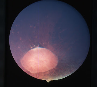
A 35 year old female presented with sudden painless decrease of vision in her right eye. Visual acuity in her right eye was 6/12 on distant Snellen’s acuity chart. A relative afferent pupillary defect was noted. Fundus examination in right eye showed optic disc swelling, retinal opacification and retinal edema with a perfused area of retina (Figure 1, arrowhead) at posterior pole suggestive of central retinal artery occlusion with patent cilioretinal artery.

central retinal artery occlusion journal
A detailed medical history revealed that she had taken a dose of depot progesterone three months back for contraception. Complete haemogram, coagulation profile and lipid profile was normal. Carotid Doppler showed presence of a thrombus in right internal carotid artery for which she is under care of cardiologist. Central retinal artery occlusion with a patent cilioretinal artery presents with constriction of visual fields but central vision is preserved.

Saturday 26 November 2016

Addressing Keratoconus disorder using as permeable lenses

GP Contact Lenses

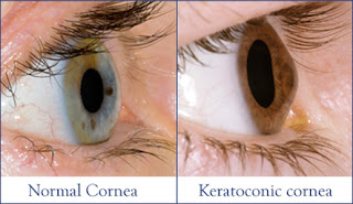
Keratoconus is a progressive corneal disorder resulting in thinning and stiffening of the central and Para central cornea. This would lead to protrusion. Although exact cause of this disorder is not known yet, genetical and environmental causes are considered responsible in causing this corneal disease. Keratoconus can be managed with spectacles and contact lenses in the early stages and it may deman surgical intervention in the advanced stage. However, corneal stability and rehabilitation can be achieved using gas permeable lenses.

Friday 25 November 2016

Management of Epiphora

optometry journals impact factor
Epiphora is a common presenting problem in ophthalmological clinical practice. There are multiple causes for epiphora. Canalicular obstruction/stenosis is common and increases with age with a female preponderance. In this first case series which used Mini Monoka stents to treat epiphora, there was a high success rate in improvement of epiphora. Further research is warranted to further validate its use into routine clinical practice.

Thursday 24 November 2016

Surgical Management of Glaucoma in Sturge-Weber Syndrome

journal of glaucoma impact factor
Sturge-Weber syndrome (SWS) or encephalo-trigeminal hemangiomatosis is a sporadic, mesodermal phakomatosis characterised by hemangiomas involving many parts of the body, particularly ocular, intracranial and facial cutaneous structures. Glaucoma is a common feature, with an incidence of 30%-71% in patients with Sturge-Weber syndrome. The aim of study was to evaluate the surgical outcomes in terms of intraocular pressure control, maintenance of visual acuity and complications of glaucoma surgeries, in eyes with Sturge-Weber syndrome and to evaluate risk factors leading to surgical failure.

Wednesday 23 November 2016

Ocular Surface Temperature and Tear Film Matrix Metalloproteinase-9 Concentration in Sjögren Syndrome Patients

Sjogren‘s syndrome (SS) is a systemic autoimmune disease characterized by polyglandular tissue destruction mainly affecting the salivar and lacrimal glands. Severe dry eye signs,including low tear volume, tear instability, inflammation of the ocular surface tissues and increased corneal staining are the main ocular manifestations of SS. Different diagnostic techniques have been developed to evaluate and diagnose dry eye syndrome; however, many of these test are invasive.

ophthalmology open access journals
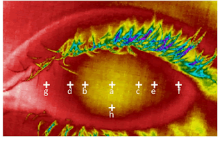
Infrared thermal images have been used in the last decades for measuring the ocular surface temperature, since its main advantage is being a non-invasive test. Another factor, which could influence the ocular surface temperature, is inflammation. o corroborate this relationship between ocular surface temperature and inflammation would be interested to measure another inflammatory molecules and ocular surface temperature in the same visit. Study was performed only in women patients with the aim to avoid any bias due to gender. Central corneal temperature in Sjögren Syndrome patients was higher than in healthy patients.

Monday 21 November 2016

Hemolytic Uremic Syndrome Complicating Invasive Streptococcus pneumoniae Infections: Tunisian Experience

Hemolytic uremic syndrome (HUS), characterized by the triad of micro-angiopathic hemolytic anemia, thrombocytopenia, and acute renal insufficiency, is a common cause of acute renal failure in children. It usually follows an episode of gastroenteritis with enterotoxigenic Escherichia coli and is termed typical HUS. However, HUS is also a complication of invasive pneumococcal infection. Reasons for not diagnosing this condition include the absence of a specific laboratory test, the lack of consistent case definitions, unfamiliarity, a misdiagnosis of disseminated intravascular coagulation (DIC), and cases with micro-angiopathic hemolytic anemia and only mild renal injury.

Invasive Streptococcus pneumoniae Infections
The aim of our study is to describe the epidemiology, the treatment and the evolution of HUS after invasive pneumococcal infections in Tunisia. Cases were identified between 2008 and 2016. Infection with S. Pneumoniae was confirmed with culture of cerebrospinal fluid, pleural fluid, or blood. Eight children fulfilled our criteria for inclusion in the study. Primary patterns were fever, respiratory signs, neurological signs and uncommon patterns. Pneumonia was a presenting feature in 6 of 8 cases (75%), two patients had confirmed pneumococcal meningitis.

Thursday 17 November 2016

Mini Monoka Stent Insertion for the Management of Epiphora Secondary to Canalicular Obstruction or Stenosis

A retrospective study was performed selecting consecutive patients at Hobart Eye Surgeons, from Oct 2007 to Mar ’08 who had Mini Monoka stents inserted for epiphora secondary to confirmed obstruction/stenosis in either inferior or common canaliculus. 

Stenosis
These patients had their stents routinely removed after six weeks. There were a total of 36 eyes (31 patients) included in the study and 31 eyes (83.7%) showed subjective improvement in epiphora. One patient was lost to follow up and 5 eyes showed no improvement. In this first case series which used Mini Monoka stents to treat epiphora, there was a high success rate in improvement of epiphora. Further research is warranted to further validate its use into routine clinical practice.

Concomitant Intraocular and Orbital Space-Occupied Lesions

Orbital SpaceConcomitant intraocular and orbital space-occupied lesions are relatively rare and this is the first report stressing the underlying causes of concomitant intraocular and intra orbital lesions. In this paper, we present cases showing both intraocular and orbital soft tissue masses and highlight their clinical, pathological and imaging features. 

Based upon the findings, the presence of a lesion involving both the orbit and eye should be included when a diagnosis of retinoblastoma, idiopathic inflammatory pseudo tumor, lymphoma, and/or Sturge- Weber syndrome is proposed.

Wednesday 16 November 2016

Need for the cost-effective techniques to detect and cure open angle glaucoma in Mexico

Open Angle Glaucoma

A transversal study estimated the cost of open angle glaucoma during the first year as $1,144,611,537 USD per annum in Mexico. The study that assessed the socio-economic costs in 30 most populous places of the country suggests the need for cost-effective method that could facilitate the early detection of open angle glaucoma with visual disability.

Friday 11 November 2016

Photodynamic Therapy with Verteporfin, a Novel Treatment Option for Retinal Arterial Macro aneurysm

First described by Robertson in 1973. RAMs are acquired, localised dilatations of an arteriole within the radius of the 3rd branch of the retinal arterial tree. Current treatment options are controversial and of unproven benefit.

Retinal Arterial Macroaneurysm
We present the case of a 76-year old gentleman with progressive vision loss from a unilateral RAM who responded favourably to treatment with PDT using Verteporfin. To the best ofour knowledge, there are no previously published reports on the use of PDT in treating RAM.


In accordance with the declaration of Helsinki, the patient provided informed consent prior to participation. We certify that all applicable institutional and governmental regulations concerning the use of human volunteers were followed during this research.

Wednesday 9 November 2016

Keratoconus Management: Role of GP Contact Lenses

Keratoconus is a corneal disorder progressive characterized by thinning and steepening of the central and Para central cornea, which leads to protrusion. The exact cause of the disease remains to be fully elucidated although its etiology probably is multi factorial with a genetic predisposition to keratoconus influenced by external environmental factors (eye rubbing and atopy) with different systemic involvement. In the early stages, keratoconus can be managed with spectacles or contact lenses (CL), but when keratoconus progresses, other surgical techniques are often required.

Keratoconus
For example, anterior lamellar (dDALK) or penetrating keratoplasty (PK) permit than 3 of 4 patients achieve best-corrected visual acuity of 20/40 or better. Intra corneal ring segment(ICRS) are proposed to increase corneal stability and decrease the astigmatism a symmetry normalizing the corneal contour with slight improvement of patients’visual acuity. Corneal collagen cross-linking (CXL) is a common technique proposed to reduce disease progression with a light improvement of visual acuity (1 to 2 Snellen lines).

Monday 7 November 2016

Best treatment for Retinal arterial macro aneurysms (RAM)


Retinal Arterial Macroaneurysm

Retinal arterial macro aneurysms (RAM) are acquired hemorrhagic or exudative focal dilations of retinal arterial branches. Treatment options that are currently available didn’t have proven benefit or high success rate. Recent studies have shown that photo dynamic therapy (PDT) is highly effective and can be used as an adjunct in the management of RAM.

Saturday 5 November 2016

Challenges in the Management of Glaucoma in a Patient with Severe Ocular Surface Disease: A Case Report

Ocular surface disease (OSD) is a significant problem for glaucoma patients worldwide, who will require long-term topical treatment. The risk of OSD increases with age, with a reported prevalence of up to 60%. The severity and burden of OSD also increase with the number of topical anti-glaucoma medications used in treatment due to multiple, daily exposures of the ocular surface (OS) to toxic active compounds in the drug itself, other components of the formulation or preservatives. 

Glaucoma with Ocular Surface Disease

The presentation of OSD varies depending on its severity, but usually includes symptoms of dry eyes, allergy and distorted vision, which can be debilitating and affect quality of life. The clinical signs of the diseaseinclude tear-film instability, toxic kerato conjunctivitis, eyelid abnormalities and allergic manifestations. However, the lack of concordance between symptoms and signs often makes the diagnosis of OSD and assessment of its severity challenging.

Friday 4 November 2016

Effect of Caffeine Intake on Retinal Microsurgical Performance

To assess the effects of prior caffeine intake on tremor control and internal limiting membrane peeling proficiency of experienced retinal surgeons using a microsurgical simulator. Experienced vitreo retinal surgeons were included. On two separate days, each subject underwent a test on a level 4 anti-tremor test and internal limiting membrane peeling test on a microsurgical simulator (Eye-Si/ Series 199, VR Magic, Software 2.9, Mannheim, Germany) first with no caffeine intake and after 40 minutes of taking an oral dose of caffeine (200 mg and 400 mg). 

Retinal Microsurgical Performance
Each subject underwent blood pressure and heart rate measurements before and 40 min after caffeine intake. The same technician measured both surgical performances. Mean age was 46.4 ± 10.1 years. All subjects were male. The mean anti-tremor results were: baseline scores 61.2 ± 19.15, 200 mg 61.6 ± 12.63 and 400 mg 75.4 ± 15.09. The mean internal limiting membrane peeling results were: baseline score 55.9 ± 5.46, 200 mg 54.8 ± 10.05 and 400 mg 62.6 ± 9.63. Blood pressure and heart rate remained stable. 

Thursday 3 November 2016

Contact Lenses: Out with the Old/In with the New?


To answer this question: I certainly hope not, at least not always. Without a doubt over the past several years the contact lens industry has brought forth numerous new materials, designs, and modes of contact lens manufacturing. But that should not translate into a universal switch from the “old” to the “new”. I would like to share some specific instances where older lens materials and designs should still have a place in your practice.


Contact LensesAlthough without a question newer more oxygen permeable rigid gas permeable (RGP) materials have been key to the introduction of such modalities as overnight wear of Orthokeratology lenses, these materials often have some less advantageous attributes. Although we often only consider oxygen permeability (Dk) and lens wettability when selecting a RGP material, one should also consider that such materials often have lower material hardness and greater modulus. 

Wednesday 2 November 2016

Superior versus Inferior Conjunctival Autograft Using Fibrin Glue in Management of Primary Pterygium: Clinical and Histopathological Results

Pterygium is a fibrovascular growth of the conjunctiva extending across the limbus to invade the cornea at a variable distance. The exposure to ultraviolet rays is a presumed a etiology and hence it is more common in people in countries with hot cimate.

Autograft Using Fibrin Glue
Surgical removal of the pterygium is the permenant treatment; however recurrence is the main issue. Simple excision leads to high rates of recurrence which are reported to be as high as 89%. Free conjunctival autograft which was first described by Kenyon et al. shows a low recurrence rate of 5.3%. Since then, many subsequent studies have found this surgical technique to be safe and effective in reducing the number of recurrences after pterygium surgery.