Monday 31 October 2016

Automatic Measurement of Choroidal Thickness with Swept-Source Optical Coherence Tomography for Clinical Follow-Up in Acute Vogt-Koyanagi-Harada Disease

Optical Coherence Tomography

Vogt-Koyanagi-Harada (VKH) syndrome is a bilateral granulomatous uveitis that typically presents with distinct clinical features based on the duration and stage of the disease. The acute stage of VKH is characterized by diffuse choroiditis, multifocal areas of subretinal fluid and/or bullous serous retinal detachments, with or without neurologic (headaches, meningismus) or auditory (tinnitus, hypoacusia) symptoms. 

The convalescent stage of the disease develops 12 weeks after onset and is characterized by resolution of retinal detachments with disappearance of cells from the anterior chamber and the vitreous, with characteristic pigmentary changes in the macula and sunset glow fundus. The chronic/recurrent phase is characterized by clinical signs of disease activity in the anterior segment of the eye with anterior granulomatous uveitis and dermatologic signs (vitiligo, alopecia, poliosis).

Thursday 27 October 2016

Photodynamic Therapy with Verteporfin, a Novel Treatment Option for Retinal Arterial Macroaneurysm

Photodynamic Therapy with Verteporfin
First described by Robertson in 1973. RAMs are acquired, localised dilatations of an arteriole within the radius of the 3rd branch of the retinal arterial tree. Current treatment options are controversial and of unproven benefit. We present the case of a 76-year old gentleman with progressive vision loss from a unilateral RAM who responded favourably to treatment with PDT using Verteporfin. To the best of our knowledge, there are no previously published reports on the use of PDT in treating RAM. 

In accordance with the declaration of Helsinki, the patient provided informed consent prior to participation. We certify that all applicable institutional and governmental regulations concerning the use of human volunteers were followed during this research.

Tuesday 25 October 2016

Aqueous Humor Outflow Structure and Function Imaging At the Bench and Bedside: A Review

Aqueous Humor Outflow Structure
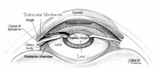
Anterior segment glaucoma clinical care and research has recently gained new focus because of novel imaging modalities and the advent of angle-based surgical treatments. Traditional investigation drawn to the trabecular mesh work now emphasizes the entire conventional aqueous humor outflow (AHO) pathway from the anterior chamber to the episcleral vein. AHO investigation can be divided into structural and functional assessments using different methods. 

The historical basis for studying the anterior segment of the eye and AHO in glaucoma is discussed. Structural studies of AHO are reviewed and include traditional pathological approaches to modern tools such as multi-model two-photon microscopy and optical coherence tomography. Functional assessment focuses on visualizing AHO itself through a variety of non-real-time and real-time techniques such as aqueous angiography. Implications of distal outflow resistance and segmental AHO are discussed with an emphasis on melding bench-side research to viable clinical applications. 

Monday 24 October 2016

Ischaemic Optic Neuropathy - Arteritic

Ischaemic Optic Neuropathy
Ischaemic optic neuropathy (ION) is the commonest adult optic nerve disorder encountered worldwide and can be expected to increase in incidence in our ageing population. The condition has been classified as a) anterior (AION) affecting the optic nerve head and b) posterior (PION) involving that portion of the optic nerve behind its immediate retrolaminar portion. Furthermore there are two pathological varieties of the disease c) Arteritic (AAION) almost exclusively associated with Giant Cell Arteritis (GCA) and d) Non arteritic (NA-AION or less correctly NAION) usually associated with diabetes, hypertension and hyper cholesterol laemia. A recent treatise on the subject runs to more than 600 pages.

Friday 21 October 2016

These are the proteins in humans that are associated with Cross-Linked Actin Networks in Trabecular Meshwork Cells

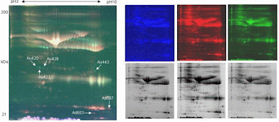
2D-DIGE

The majority of the aqueous humor, which is produced by the ciliary body, is drained through the conventional outflow pathway. In this pathway, the aqueous humor flows from the posterior chamber to anterior chamber and exits the eye through the trabecular mesh work(TM), a multilayered tissue in the iridocorneal angle of the eye.


In primary open angle glaucoma (POAG), glucocorticoid-induced ocular hypertension (GC-OHT), and glucocorticoid induced glaucoma (GIG), the outflow resistance at the TM is increased, which causes intraocular pressure (IOP) elevation. Elevated IOP is the major risk factor for the development and progression of glaucoma that leads to irreversible visual loss that can cause blindness. 

Thursday 20 October 2016

Can coffee intake affect the Retinal Microsurgical Performance?

Retinal Microsurgical

Coffee is one of the most widely consumed nonalcoholic beverages in the world. Caffeine in coffee acts on central nervous system through A1 and A2 subtypes of the adenosine receptors that help in increasing alertness, vigilance and reducing fatigue. However Caffeine consumption is highly discouraged prior to Retinal Microsurgery by micro surgeons as caffeine can cause potential adverse effects.

Monday 17 October 2016

Aqueous Humor Outflow Structure and Function Imaging At the Bench and Bedside: A Review

Anterior segment glaucoma clinical care and research has recently gained new focus because of novel imaging modalities and the advent of angle-based surgical treatments. Traditional investigation drawn to the trabecular mesh work now emphasizes the entire conventional aqueous humor outflow (AHO) pathway from the anterior chamber to the episcleral vein. AHO investigation can be divided into structural and functional assessments using different methods. 

Aqueous Humor Outflow Structure
The historical basis for studying the anterior segment of the eye and AHO in glaucoma is discussed. Structural studies of AHO are reviewed and include traditional pathological approaches to modern tools such as multi-model two-photon microscopy and optical coherence tomography. Functional assessment focuses on visualizing AHO itself through a variety of non-real-time and real-time techniques such as aqueous angiography. Implications of distal outflow resistance and segmental AHO are discussed with an emphasis on melding bench-side research to viable clinical applications. 

Friday 14 October 2016

Paediatric Aphakic Glaucoma: A Diagnostic and Management Challenge

Pediatric aphakic glaucoma is a potentially blinding condition, which poses significant management challenges - the patient is young and treatment must aim to preserve vision for perhaps 80 years or more of life expectancy. The purpose of this study is to highlight that early detection is important to improve the prognosis for these patients and their families. Misdiagnosis of this condition may adversely affect vision preservation. 

Paediatric Aphakic Glaucoma
However, diagnosis can be difficult, especially when children may demonstrate vague symptoms such as irritability, photophobia and epiphora which themselves may further impair clinical co-operation. Despite considerable advances in techniques employed in paediatric cataract surgery, aphakic glaucoma continues to occur and remains notoriously challenging to manage. We present a case of glaucoma after cataract surgery that masqueraded as corneal infection which was challenging to diagnose and treat. Prompt recognition of pediatric aphakic glaucoma and intraocular pressure (IOP) control has resulted in a favorable clinical outcome, thus far.

Thursday 13 October 2016

Fundus Auto fluorescence and Enhanced Depth Imaging Spectral-Domain Optical Coherence Tomography in Hunter Syndrome-New Insights

Hunter syndrome or mucopolysaccharidosis type II is a rare progressive multi-systemic disorder, caused by an abnormal storage of glycosaminoglycans (GAGs) in almost every cell type, including most ocular tissues. Patients have a short life expectancy and ocular manifestations can be present early in the course of disease. To report the fund us auto fluorescence and tomographic ocular findings in Hunter syndrome. A 18-year-old male patient with Hunter syndrome with progressive nyctalopia was submitted to color fundus photography, blue fundus auto fluorescence (FAF), fluorescein angiography (FA) and spectral domain optical coherence tomography with enhanced-depth imaging (EDI-SD OCT). 

Spectral-Domain Optical Coherence Tomography
Results and discussion: Fundus examination and wide-field fluorescein angiogram revealed normal optic discs and bilateral pigmentary atrophic changes at the mid periphery with macular sparing. SD OCT revealed a retinal thinning due to external retinal atrophy affecting the photoreceptor layer beyond the parafoveal area. Although a prominent central external limiting membrane (ELM) was present, both the ellipsoid zone band and ELM could not be tracked beyond the central 2-mm and 2.5 mm diameter ring, respectively. EDI-SD OCT revealed a highly irregular choroid, especially in its outer boundary, probably due to GAG scleral deposition. Blue FAF presented a symmetric hyper auto fluorescent parafoveal ring that corresponded to the area where the ELM was present in the absence of the ellipsoid band.

Wednesday 12 October 2016

Impact of Amblyopia Treatment on Vision-Related Quality Of Life

Treatment of amblyopia during childhood can have both positive and negative impacts in later life. The treatment of amblyopia could influence Vision-related Quality of Life in adults and children, particularly social relationships and emotions. For example, children with a history of occlusion were 35% to 37% more likely than children without visual defects to have suffered from verbal or physical bullying at school. Many parents associate occlusion treatment with a decrease in children’s self-confidence because of poor vision during occlusion and report greater distress and more conflict at home.

Amblyopia Treatment
Koklanis et al. conducted a study in Australia on the psychosocial impact of amblyopia and its treatment from both the children’s (aged 3 to 15 years) and parents’ perspectives. The investigators performed a semi-structured interview with both children with amblyopia andtheir parents. In addition, parents were asked to complete a psychological inventory, the Behaviour Assessment System for Children. 

Monday 10 October 2016

Prologue to the Special Issue: Age-Related Macular Degeneration

Age-related macular degeneration (AMD) is a leading cause of irreversible vision loss among people age 50 and older. Based on the presence or absence of blood vessels, it is classified intotwo types, wet and dry. In the past decades, significant progress has been made in understanding the molecular mechanisms underlying wet AMD, and several robust therapeutic drugs were developed to block the development of new blood vessels and leakage from the abnormal vessels with favorable clinical effect.

Macular Degeneration
In contrast, there are no approved treatments for dry AMD and the mechanisms are not completely known. However, it has been approved that genetics, complement dysregulation, oxidative stress, mitochondria DNA damage were involved in the mechanisms. Variety of scientific studies, including gene replacement therapy, retinal cell transplantation, pharmaceutical intervention and vitamin dietary supplementation, hold promise in developing treatment to prevent or slow the progression of the disease. In the clinical aspect, multiple clinical studies and trials have been done to further our knowledge of AMD, and on-going studies are raising hopes for improved treatments.

Friday 7 October 2016

Balancing Patient and Practitioner Goals in Contact Lens Fitting

Patients generally are seeking three things from their contact lens wearing experience, vision, comfort, and convenience. In addition, some individuals will be looking to seek these goals with a minimum of “out of pocket” expense. As an eye care provider our goal incontact lens fitting is to provide for optimal ocular health while also establishing a vision correction modality that maximizes clear binocular vision. However, there may be situations when we need to strike a balance between what patients can and will do, as contrasted with what might otherwise seem the ideal situation.

Contact Lens Fitting
There is a wealth of published data that clearly indicates that patients do not always replace their lenses with the frequency we would like. Sometimes this is due to negligence, but often the underlying motivation is simply cost. We need to remind ourselves that the replacement period for most contact lenses is not dictated by the FDA, but is based on recommendations made by the manufacturer. 

Thursday 6 October 2016

Bilateral Chronic Central Serous Chorioretinopathy (CSCR) Induced by Long-Term Testosterone Treatment

Purpose: We reported a rare case of bilateral chronic central serous chorioretinopathy (CSCR) induced by longterm exogenous testosterone treatment. Method: A case report. Result: A 52-year-oldman with medical history of diabetes mellitus, hypertension, diabetes insipidus and hypogonadism presented to our ophthalmologic clinic with unstable blurred vision of right eye for more than 5 years and vision loss of left eye since childhood injury. Optical coherence tomography (OCT) showed subfoveal fluid in both of his eyes. 

Chorioretinopathy
Fluorescein angiography (FA) revealed fluorescein in multiple leaking points around the arcade area, which appeared hyper fluorescence not only in early phase but sustained fluorescein pooling till late phase in both eyes. According to his medical records, he received intramuscular testosterone injection every two weeks as treatment of hypogonadism for more than 10 years. His serum level of testosterone was higher than normal range. After focal photocoagulation in his right eye combined with decreasing frequency of testosterone intramuscular injection, the subfoveal fluid in both of his eyes totally resolved with vision improvement.